


Case Report: What’s Your Diagnosis?
Author: Vivian Wong MD-PhD, Department of Dermatology, Warren Alpert Medical School of Brown University; Providence, Rhode Island
Case history:
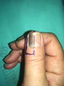
A 34-year-old female (Fitzpatrick Skin Phototype V) presents with 8-month history of nail discoloration on her right thumb. On examination, there is a 4.3 mm brown patch composed of irregular brownish to grayish longitudinal lines of various widths on the medial nail plate. The lesion does not disappear with pressure. The other finger and toe nails are uninvolved. She denies any preceding trauma and does not take any medication. What is your diagnosis?
- Subungual hematoma
- Subungual melanoma
- Physiologic pigmentation
- Pigmented onychomycosis
Answer:
- Subungual melanoma
Explanation:
Subungual melanoma is a type of melanoma involving the nail apparatus. It has a predilection for patients with darker skin phototypes. Subungual melanoma is a diagnostic challenge, as numerous conditions could present with nail pigmentation. Several common culprits are discussed here. In addition, dermatologists should always obtain a thorough history to rule out exogenous or medication-induced melanonychia. Therefore, the diagnosis is often delayed, resulting in significant mortality. Further, amelanotic subungual melanoma has been reported, and it could mimick other neoplastic or inflammatory processes, such as squamous cell carcinoma, pyogenic granuloma, and viral warts.
As in this case, subungual melanoma favors a single digit, most likely the thumb of an adult. Examination of the lesion using dermoscopy is often helpful. Signs suggestive of the diagnosis include irregular pigmentation, broad pigment band (at least 3mm), tapered band with a wider base at the origin of the nail plate, blurry and fuzzy borders, and variegated color. Hutchinson’s sign, with pigmentation of periungual tissue, is a classic finding. Laugier-Hunziker syndrome, a benign hereditary pigmentary condition, could present with benign nail pigmentation. Perform serial photography for monitoring if suspicion is low, and perform a nail biopsy if suspicion is high.
Differential diagnoses:
-
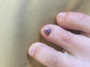
- Subungual hematoma refers to a collection of blood within the nail plate or nail bed, often secondary to trauma. It could resemble subungual melanoma, with red-brown to brown-black discoloration. It typically migrates distally and spontaneously resolves with time, unlike subungual melanoma.
- Subungual hematoma refers to a collection of blood within the nail plate or nail bed, often secondary to trauma. It could resemble subungual melanoma, with red-brown to brown-black discoloration. It typically migrates distally and spontaneously resolves with time, unlike subungual melanoma.
-
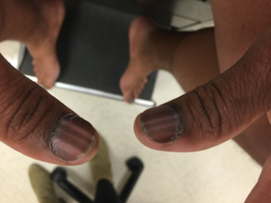
- Physiologic (functional pigmentation) is typically seen in patients of darker skin phototypes. A clue to diagnosis is the involvement of multiple nails. Look for regular longitudinal and rectangular pigment bands with homogenous color.
- Physiologic (functional pigmentation) is typically seen in patients of darker skin phototypes. A clue to diagnosis is the involvement of multiple nails. Look for regular longitudinal and rectangular pigment bands with homogenous color.
-
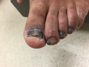
- Pigmented onychomycosis (fungal melanonychia) occurs secondary to dematiaceous fungal or rarely yeast and mold infections of the nails. Look for subungual debris, nail thickening, yellowish discoloration and onycholysis, in addition to brown-black discoloration. Multiple nails, typically the toe nails, are involved. Looks for evidence of concomitant tinea pedis, which presents with scaly plaques on the soles, lateral aspects of the feet and the interdigital spaces.
- Pigmented onychomycosis (fungal melanonychia) occurs secondary to dematiaceous fungal or rarely yeast and mold infections of the nails. Look for subungual debris, nail thickening, yellowish discoloration and onycholysis, in addition to brown-black discoloration. Multiple nails, typically the toe nails, are involved. Looks for evidence of concomitant tinea pedis, which presents with scaly plaques on the soles, lateral aspects of the feet and the interdigital spaces.
References:
Mun, J.-H., Kim, G.-W., Jwa, S.-W., Song, M., Kim, H.-S., Ko, H.-C., Kim, B.-S. and Kim, M.-B. (2013), Dermoscopy of subungual haemorrhage: its usefulness in differential diagnosis from nail-unit melanoma. British Journal of Dermatology, 168: 1224–1229.
Braun RP, Baran R, Le Gal FA, Dalle S, Ronger S, Pandolfi R, et al. Diagnosis and management of nail pigmentations. J Am Acad Dermatol. 2007;56:835–847.
Dominguez-Cherit, J., R. Roldan-Marin, P. Pichardo-Velazquez, C. Valente, V. Fonte-Avalos, M. E. Vega-Memije, and S. Toussaint-Caire. “Melanonychia, Melanocytic Hyperplasia, and Nail Melanoma in a Hispanic Population.” [In Eng]. J Am Acad Dermatol 59, no. 5 (Nov 2008): 785-91.
Wong V, Burgin S, Richert B, Baran R. “Subungual melanoma (nail melanoma)”. In: Goldsmith LA, editor. VisualDx. Rochester, NY: Logical Images. https://www.visualdx.com/visualdx/diagnosis/subungual-melanoma?moduleId=19&diagnosisId=56099
Wong V, Burgin S, Richert B, Baran R. “Onychopapilloma”. In: Goldsmith LA, editor. VisualDx. Rochester, NY: Logical Images. https://www.visualdx.com/visualdx/diagnosis/onychopapilloma?moduleId=19&diagnosisId=56100

Cutis Journal
Read published peer-reviewed articles written your by Skin of Color Society members
Join Our Newsletter
Stay up-to-date on the latest news and happenings related to The Skin of Color Society.

Did You Know
Skin of color patients comprise the majority in California, New Mexico and Texas…and soon will be the majority in Arizona, Nevada, Georgia, New York and Florida.
By 2042, more than 50% of the US population will have skin of color.